Histopathology & Biopsy Lab in Vijayanagar, Bangalore
Padmashree Diagnostics runs a dedicated biopsy testing lab in Vijayanagar, Bangalore. Our Department of Histopathology studies tissue specimens to give you an accurate diagnosis. This is the foundation of good medical treatment.
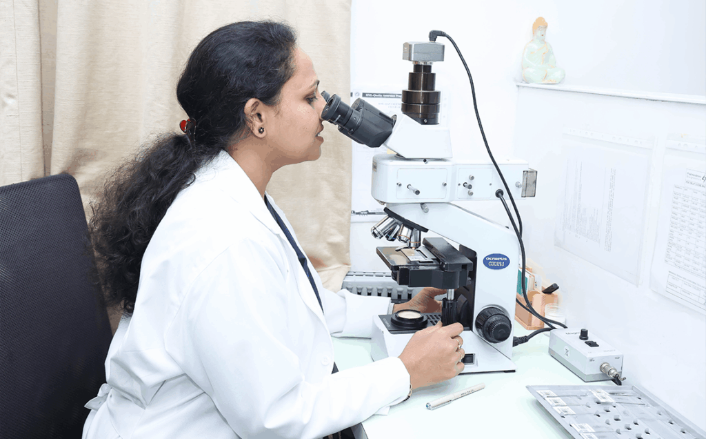
We study cells and tissues under the microscope. This helps doctors diagnose diseases and check how severe they are. It also guides treatment decisions and helps track patient outcomes over time.
We process every sample with care. This includes routine biopsies and complex cancer diagnosis cases alike. Our experienced consultant pathologists interpret each sample. This helps your doctor choose the right treatment with confidence.
What Is Histopathology?
Histopathology is the study of diseased tissue under a microscope. Doctors usually collect this tissue through a biopsy or a surgical procedure.
Cytology looks at individual cells alone, often collected from bodily fluids or a fine needle sample. Histopathology is different. It keeps the tissue’s structure intact. This lets pathologists see how cells relate to the tissue around them.
This difference matters for your treatment. Histopathology often confirms what an earlier FNAC or scan first suggested. In most cases, it gives the clearest and most final diagnosis before treatment planning begins.
From Biopsy to Diagnosis: Our Process
Good tissue diagnosis starts with careful sample handling. Our lab uses advanced Leica equipment. This keeps tissue processing consistent and produces clear microscopic slides.
Pathologic examination starts with a gross description. Our team looks at, measures, and records the size, color, and texture of each tissue specimen before any cutting begins. Technicians then prepare thin histologic sections from the tissue and mount them on a glass slide for the pathologist to study.
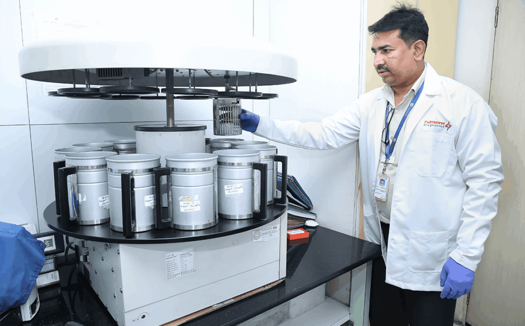
Leica Automated Tissue Processor
This machine processes every tissue sample the same way, every time. Consistent handling lowers the risk of errors that could affect your diagnosis.
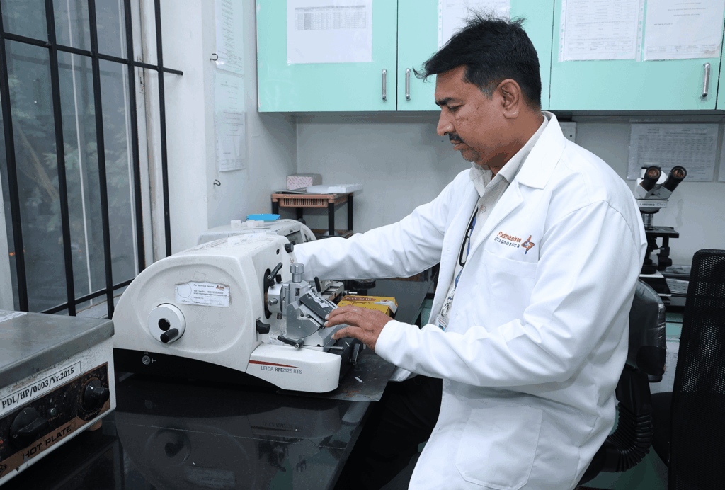
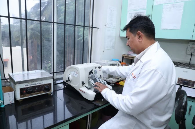
Leica Rotary Microtome
Histo technologists use this equipment to cut very thin, precise slices of tissue. These thin slices are needed for microscopic study. This step is critical. It produces slides a pathologist can read with confidence.
Molecular Pathology and Advanced Testing
Some cancer cases need more than a microscope alone. In these cases, we draw on molecular pathology. A technique called fluorescence in situ hybridization (FISH) lets pathologists study the genetic makeup of cancerous cells in close detail. This can help doctors choose more targeted, effective treatment options.
Biopsy and Surgical Pathology Services We Support
Our histopathology department handles a wide range of biopsy and surgical pathology cases. This includes samples from the skin, breast, gastrointestinal tract, gynaecological system, and lymph nodes. We also handle many other types of surgical specimens.
For some complex cases, we apply special stains. These stains highlight specific cells or structures. This extra step helps our pathologists reach a clear diagnosis in tricky cases.
When Might Your Doctor Recommend a Biopsy?
Your doctor may recommend a biopsy in a few common situations. First, imaging tests like ultrasound, mammography, or a CT/MRI scan may find a lump or unusual growth. Second, a physical exam may find something that needs a closer look. In both cases, a biopsy gives a clear answer about what the tissue actually is. Doctors also order a biopsy after surgery. This confirms exactly what type of tissue the surgeon removed.
A biopsy is a normal, well-established step in diagnosis. On its own, it does not mean you have cancer.
Preparing for a Biopsy Procedure
What Padmashree Diagnostics Does for Biopsy Testing
Padmashree Diagnostics does not perform the biopsy procedure itself. Hospitals and clinicians across Bangalore refer biopsy specimens to our laboratory, where our pathology team carries out detailed histopathological examination and reports the findings back to the referring hospital or doctor.
Our process includes:
Receiving specimens with proper documentation – each specimen is accepted along with a duly filled requisition form from the referring doctor, specifying clinical history and biopsy site.
Standardized processing and examination – specimens are processed, sectioned, stained, and examined under strict laboratory protocols to ensure accurate and consistent results.
Correlation with clinical history – where available, relevant clinical history and prior reports are reviewed to support accurate interpretation of findings.
Verified reporting – findings are reviewed by our pathologists before the final report is issued and sent back to the referring hospital or doctor for discussion with the patient.
Turnaround Time and Getting Your Report
Histopathology takes more steps than a simple blood test. We process, section, and stain the tissue before a pathologist reviews it. Because of this, reports take a bit longer than blood or urine tests. Most reports are ready within a few working days. The exact time depends on how complex your case is. Cases that need special stains may take a little longer.
Our team prepares detailed pathology reports for every case, so your doctor gets the full clinical picture, not just a one-line result. We will confirm a timeline once we receive your sample. You can then download your report through our online Lab Reports portal as soon as it is ready.
Book Your Histopathology / Biopsy Test in Vijayanagar, Bangalore
If your doctor has advised a biopsy, you can drop off your sample at our Vijayanagar lab. Lab reports can be collected by walking to the center as the reports will be issued along with the slides.
Why Choose Padmashree Diagnostics for Histopathology?
Experienced consultant histopathologists reviewing every case
Advanced Leica Automated Tissue Processor for consistent sample quality
Precision Leica Rotary Microtome for high-quality microscopic sections
Frozen section analysis available for urgent surgical cases
Special stains available for selected complex cases
NABL-aligned quality standards across the department
Timely reporting with expert, clinically correlated interpretation
List of Histopathology Tests
Showing 0 of 0 services
1Biopsy
Microscopic examination of tissue sample to diagnose disease, identify malignancy, assess inflammation, and guide clinical management decisions accurately and treatment.
2Biopsy Extra Large Specimen
Detailed histological analysis of extra large tissue specimens to diagnose complex diseases, evaluate margins, staging, and guide treatment planning accurately.
3Biopsy LargeSpecimen -1
Histopathological examination of large tissue specimen for disease diagnosis, tumor grading, margin assessment, and correlation with clinical findings accurately reported.
4Biopsy Large Specimen -2
Comprehensive microscopic evaluation of large biopsy specimen to detect pathology, assess tumor extent, margins, and support definitive diagnosis accurately clinically.
5Biopsy MediumSpecimen -1
Microscopic analysis of medium-sized tissue specimen to identify disease processes, inflammation, neoplasia, and assist appropriate clinical management decisions accurately promptly.
6Biopsy Medium Specimen -2
Histopathology evaluation of medium biopsy sample for diagnosing lesions, grading tumors, assessing margins, and correlating with clinical information accurately documented.
7Biopsy Medium Specimen -3
Detailed tissue examination of medium biopsy specimen to confirm diagnosis, evaluate disease severity, and support treatment planning decisions accurately clinically.
8Biopsy Small Specimen
Microscopic assessment of small tissue specimen to detect pathological changes, early malignancy, infections, or inflammatory conditions accurately diagnosed and reported.
9Cell Block
Cell block preparation concentrates cellular material from fluids for histological examination, improving diagnostic accuracy and enabling ancillary testing techniques effectively.
10Slide Review for Second Opinion 2 slides
Independent review of two histopathology slides by expert pathologist to confirm diagnosis and ensure diagnostic accuracy consistency, reliability, confidence clinically.
11Slide Review for Second Opinion (Many Slides)
Comprehensive second opinion review of multiple histopathology slides to reassess diagnosis, staging, and pathological interpretation accuracy ensuring clinical confidence outcomes.
12Slide Review for Second Opinion (Per Slide)
Expert pathological review conducted per slide to verify findings, clarify diagnosis, and enhance reporting accuracy consistency, reliability, confidence clinically overall.
13Slide Review for Second Opinion with IHC Panel
Second opinion slide review combined with immunohistochemistry panel to refine diagnosis, classify tumors, and improve diagnostic precision accuracy confidence clinically.
Showing 0 of 0 tests
Frequently Asked Questions
What is the difference between a biopsy and FNAC?
FNAC uses a thin needle to collect a small number of cells. A biopsy, on the other hand, removes a small piece of whole tissue and keeps its structure intact. This usually gives a more detailed and more final diagnosis than FNAC alone.
How long does a histopathology report take?
Most biopsy reports are ready within 72-96 hrs depending upon the size of the specimen. Complex cases that need special stains may take a bit longer. We will confirm the exact timing once we receive your sample.
Does a biopsy always mean doctors suspect cancer?
No. A biopsy is a routine diagnostic step. Doctors use it to identify many types of tissue changes. Most of these changes turn out to be harmless or caused by inflammation, not cancer.
Who examines my tissue sample?
An experienced consultant histopathologist examines every sample by hand. They compare the microscopic findings with your clinical history to give you an accurate, meaningful report.
Is Padmashree's histopathology department NABL accredited?
Yes. Our histopathology processes follow NABL-aligned quality standards. We back this up with advanced Leica tissue-processing equipment and consistent laboratory protocols at every step.